
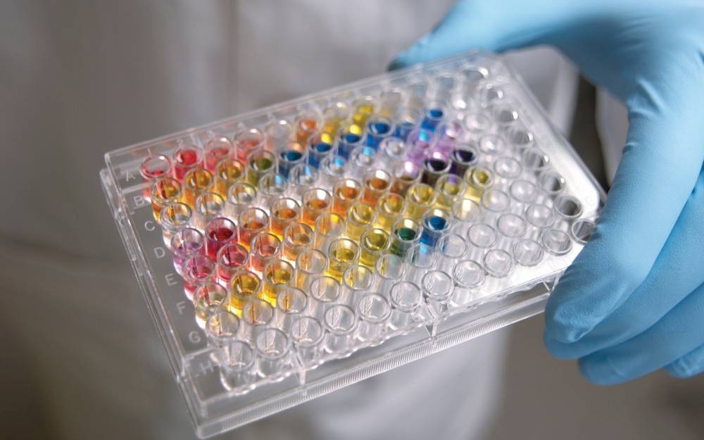
In the world of cell biology, calcium isn’t just a mineral that makes your bones strong. It is the ultimate messenger, a universal signal that tells cells when to move, when to grow, and even when it is time to die. If you think of a cell as a complex city, calcium ions are the electrical pulses that keep the lights on and the traffic flowing. Because calcium is so central to everything a cell does, scientists have spent decades perfecting ways to measure it. This is where the calcium assay comes into play, serving as a vital window into the inner workings of living organisms.
When we talk about a calcium assay, we are describing a set of techniques used to monitor the concentration of calcium ions within a cell or a biological sample. Most of the time, we are interested in intracellular calcium. Under normal conditions, the concentration of calcium inside a cell is kept incredibly low, while the concentration outside is much higher. When a cell is stimulated—perhaps by a hormone, a neurotransmitter, or a drug—calcium channels open, and a sudden surge of ions enters the cytoplasm. This transient spike is what triggers a biological response, and being able to see and measure that spike is crucial for modern medicine and drug development.

The mechanics of measuring calcium ions
You might wonder how we actually ‘see’ something as small as an ion. Since calcium ions don’t have a natural colour or shine, we have to use molecular tools that act as sensors. Most modern assays rely on fluorescence. These sensors are molecules designed to change their light-emitting properties when they bind to calcium. When the sensor is ’empty,’ it might glow dimly; when it catches a calcium ion, it shines brightly or changes its colour entirely.
There are a few different ways to get these sensors into the cells we want to study:
- Fluorescent Dyes: These are chemical compounds like Fura-2 or Fluo-4. They are often modified with ‘acetoxymethyl’ (AM) groups that allow them to slip through the cell membrane. Once inside, the cell’s own enzymes chop off the AM group, trapping the dye inside the cell where it can wait for a calcium signal.
- Genetically Encoded Calcium Indicators (GECIs): These are proteins, such as GCaMP, that cells are engineered to produce themselves. This is particularly useful for long-term studies or for looking at specific types of cells within a living tissue.
- Bioluminescent Proteins: Some assays use proteins like aequorin, derived from jellyfish. Instead of needing an external light source to glow, these proteins produce light naturally when they react with calcium.

Mia Turner is a dedicated gaming journalist passionate about exploring gameplay mechanics, esports, and community-driven content. Her work highlights tips, trends, and player insights, aiming to engage and inform both casual and hardcore gamers.